


Discover Our Impact
Device Impact
A user familiar with PI-RADS and the significance of such data will be able to view and interpret this additional information during the diagnosis of prostate cancer and improve his/her performance for interpreting both true positive and true negative suspect regions.
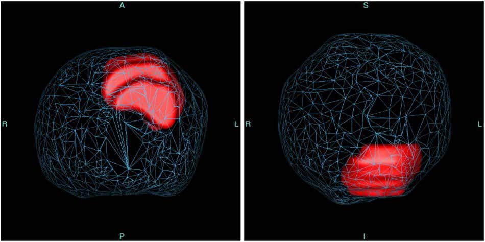
Additionally, the colorized 2D outputs of the software lend themselves to be excellent discussion graphics between physician and patient. The wholistic views of the entire prostate organ also provides the physician an excellent planning tool for additional care. The 3D output provides axial and coronol views of a translucent prostate with individual risk-color-coded lesions within. These provide excellent guidance for cognitive biopsies.
Impact on the Patient
Users should utilize all available MR data consistent with ACR clinical recommendations (e.g., dynamic contract enhanced images if available) in context of PI-RADS v2, and in conjunction with bi-parametric MRI acquired with either surface or endorectal MRI accessory coils from compatible MRI systems to make the diagnosis. Benefits to the patient could be reduction or elimination of unnecessary biopsies of non-cancerous tissue and/or more accurate or early detection of prostate cancer.

Renowned Urologist Endorses ProstatID as Game-Changer in Prostate Health Assessment
Let me begin by conveying my sincere amazement with ProstatID: I am thoroughly astounded by its capabilities. It’s remarkable efficiency in providing quick results has greatly facilitated my practice as a urologist. ProstatID transcends the limitations of conventional radiology reporting by offering a visual understanding of prostate conditions, thereby enhancing communication with patients and guiding diagnostic and treatment decisions with exceptional precision.
My experience with ProstatID has demonstrated its profound impact. On numerous occasions, I have encountered disparities between ProstatID’s findings and conventional radiological assessments. Upon reviewing cases of patients with multiple negative MRIs but persistent clinical symptoms suggestive of underlying cancer, ProstatID revealed previously undetected cancerous lesions. This discovery has been instrumental in guiding targeted biopsies and therapeutic interventions, ultimately leading to more accurate diagnoses and improved patient outcomes.
Furthermore, through retrospective analysis, ProstatID has unveiled a concerning trend in the progression of lesions from minor suspicion to significant pathology over time. This invaluable insight has informed proactive management strategies, allowing for early intervention and prevention of disease progression.
In my practice, I have seamlessly integrated ProstatID’s 3D outputs into cognitive biopsies, surpassing the limitations of traditional fusion systems. The precision and clarity provided by ProstatID’s axial and coronal views have significantly enhanced biopsy procedures, ensuring precise alignment and optimal treatment guidance.
Without a doubt, every urologist dealing with prostate conditions should incorporate ProstatID into their practice. Its indispensable role in improving diagnostic accuracy and guiding therapeutic interventions makes it an indispensable tool in the realm of urology. To forego the utilization of ProstatID would be a disservice to both patients and practitioners alike.
Russell Locke, M.D.
Vantage Urologic Institute
For more information or setting up a demonstration, please contact [email protected].