


Blog
The field of medical imaging is in a constant state of evolution, and nowhere is this more apparent than in prostate MRI interpretation. For decades, the radiologist’s trained eye was the sole authority in identifying suspicious lesions. Today, the landscape includes a powerful new ally: artificial intelligence. The introduction of AI has shifted the conversation from human-only reads to a new era of AI-augmented decision-making. This raises a critical question: how do these sophisticated algorithms compare to experienced radiologists when it comes to detecting and classifying prostate lesions? The answer is not about competition, but collaboration.
The Changing Role of Radiologists in Prostate MRI
The radiologist’s role in diagnosing prostate cancer has transformed significantly. What was once a practice centered on anatomical review has become a data-rich discipline, where AI-powered tools offer deeper insights and support clinical judgment in ways previously not possible.
From anatomy-based reading to data-driven insights
Historically, interpreting a prostate MRI involved a meticulous visual assessment. Radiologists would examine various image sequences, looking for anatomical changes, signal abnormalities, and other visual cues that suggested malignancy. While effective, this approach relied heavily on individual experience and subjective interpretation.
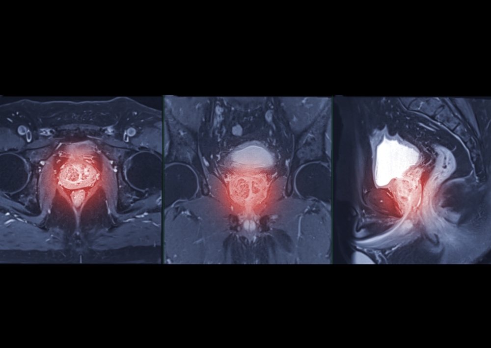
The advent of AI and radiomics—the process of extracting vast amounts of quantitative data from medical images—has added a new layer of objectivity. Instead of just looking at an image, AI systems can analyze thousands of features within a lesion, from texture and shape to intensity patterns that are invisible to the human eye. This shift moves radiology from a purely visual field to a data-driven one, where quantitative insights support and enhance the radiologist’s expert evaluation.
Challenges in human interpretation
Even for the most skilled radiologists, interpreting prostate MRI scans presents several challenges. A primary issue is inter-reader variability, where different radiologists may interpret the same scan and arrive at different conclusions. This variability can stem from experience levels, training, and the inherent subjectivity of grading systems like PI-RADS (Prostate Imaging Reporting and Data System).
Furthermore, the clinical environment is demanding. Radiologists face immense time pressure, reading numerous complex studies daily. This can increase the risk of missing subtle or early-stage lesions. The complexity of prostate anatomy itself, often complicated by conditions like benign prostatic hyperplasia (BPH) or prostatitis, adds another layer of difficulty, making it hard to distinguish cancerous tissue from benign changes.
Why AI entered the picture
AI was not developed to replace radiologists but to address these very challenges. AI-powered software provides a consistent, objective analysis every time, significantly reducing inter-reader variability. It acts as a tireless second reader, flagging suspicious areas that might otherwise be overlooked during a busy workflow. This allows radiologists to work more efficiently and focus their attention on the most critical findings.
The goal of radiology AI is to enhance human expertise by providing a safety net. It offers a powerful tool for improving consistency, boosting efficiency, and enabling earlier, more accurate lesion detection, which ultimately leads to better patient outcomes.
Measuring Performance: Radiologist vs AI
To understand how AI and radiologists compare, we must use standardized, objective measures. These metrics help quantify diagnostic accuracy and provide a clear framework for evaluating performance in clinical studies.
Key evaluation metrics
Three of the most common metrics used to measure diagnostic performance are sensitivity, specificity, and the Area Under the Curve (AUC).
- Sensitivity: This measures the ability to correctly identify patients with the disease. A tool with high sensitivity will find most true cancers. For example, if 100 cancerous lesions are present and an AI detects 95 of them, its sensitivity is 95%.
- Specificity: This measures the ability to correctly identify patients without the disease. High specificity means fewer false alarms. If 100 benign areas are scanned and a tool correctly identifies 98 as non-cancerous, its specificity is 98%.
- Area Under the Curve (AUC): This metric provides a comprehensive summary of a diagnostic tool’s overall performance across all sensitivity and specificity levels. An AUC of 1.0 represents a perfect test, while an AUC of 0.5 indicates performance no better than a random guess. A higher AUC value signifies a more accurate test.
Per-lesion and per-patient evaluation frameworks
Performance can be measured in two main ways: on a per-lesion or per-patient basis.
- Per-lesion evaluation focuses on the accuracy of identifying individual suspicious areas within the prostate. This is useful for assessing how well an AI model can pinpoint specific tumors.
- Per-patient evaluation provides a broader view, determining the overall risk for the patient as a whole. This framework assesses whether the final diagnosis for the patient—cancer or no cancer—is correct, regardless of how many individual lesions were identified. Both frameworks are essential for a complete picture of diagnostic performance.
Comparative Studies and Clinical Evidence
A growing body of research has explored the performance of AI in prostate MRI, both as a standalone tool and as an assistant to radiologists. The evidence consistently points toward a future where human-AI collaboration is the new standard of care.
AI vs expert radiologists in lesion detection
Numerous published studies have directly compared the standalone performance of AI algorithms to that of expert radiologists. In many of these studies, modern AI systems have demonstrated lesion detection accuracy that is comparable to, and in some cases superior to, human readers. These algorithms excel at identifying subtle patterns and can process vast amounts of imaging data without fatigue, giving them an edge in consistency and speed.
AI-assisted vs unaided radiologist performance
The most compelling evidence comes from studies analyzing the performance of radiologists with AI support versus those without it. This is where the true value of AI becomes clear. Research consistently shows that radiologists who use AI as a supportive tool outperform their unaided colleagues. The AI acts as a safety net, highlighting potential lesions and providing quantitative risk scores that help confirm or challenge a radiologist’s initial impression. This collaborative approach leads to higher sensitivity and greater diagnostic confidence.
Reproducibility and reader consistency
One of the most significant benefits of integrating AI into the clinical workflow is the improvement in reproducibility. AI algorithms apply the same analytical criteria to every scan, effectively eliminating the inter-reader variability that has long been a challenge in prostate MRI. By standardizing the initial analysis, AI helps ensure that a patient’s diagnosis is not dependent on which radiologist reads the scan or which institution performs the imaging. This leads to more consistent and reliable care across the board.
When Human Expertise Still Leads
Despite its power, AI is a tool, not a replacement for clinical judgment. There are many scenarios where the experience, intuition, and contextual understanding of a radiologist remain indispensable.
Complex or ambiguous cases
AI models are trained on specific datasets and may struggle with cases that fall outside their learned patterns. Radiologists are uniquely equipped to handle ambiguity. They can identify subtle imaging artifacts that might confuse an algorithm, recognize rare pathologies not included in the AI’s training data, and interpret complex presentations like multi-focal disease where multiple lesions with varying characteristics are present.
Clinical context and judgment
A radiologist’s diagnosis is never made in a vacuum. They integrate the imaging findings with a patient’s complete clinical picture, including PSA levels, biopsy history, family history, and prior imaging studies. Most AI models do not have access to this rich contextual data. This holistic view allows a radiologist to make nuanced judgments that an algorithm, focused solely on image data, cannot.
Interpretability and accountability
For a diagnosis to be actionable, the clinician must understand and trust the reasoning behind it. This is a concept known as “explainability.” While many AI models operate as “black boxes,” radiologists can articulate their diagnostic reasoning. Ultimately, the radiologist remains accountable for the final report. They must be able to verify and stand behind the AI’s findings before incorporating them into a patient’s care plan.
Hybrid Intelligence — The Best of Both Worlds
The optimal approach to prostate cancer diagnosis is not a contest between human and machine but a partnership. “Hybrid intelligence” combines the strengths of both AI and radiologists to create a system that is more accurate, efficient, and reliable than either could be alone.
Human–AI collaboration in prostate MRI
In a hybrid reading model, the workflow is designed for collaboration. The AI software first performs an automated analysis of the bpMRI scan, segmenting the prostate and highlighting any suspicious lesions with a corresponding risk score. The radiologist then reviews this AI-generated output alongside the original images. This allows them to use the AI’s findings as a guide, confirming its suggestions while applying their own expertise to make the final determination.
Improving diagnostic speed and workflow efficiency
AI significantly accelerates the reading process. By automatically flagging suspicious areas, it allows radiologists to triage cases more effectively and focus their limited time on the most clinically significant findings. This automation extends to reporting, where AI can pre-populate reports with standardized measurements and lesion data. The result is a more efficient clinical workflow, reduced turnaround times, and more capacity to handle growing patient loads.
Reducing unnecessary biopsies and false positives
One of the key goals in prostate cancer diagnosis is to avoid unnecessary invasive procedures. By improving diagnostic specificity, AI helps radiologists better distinguish between clinically significant cancer and benign conditions or low-risk disease. This increased confidence in ruling out cancer can reduce the number of false positives, sparing many men from the anxiety and potential complications of an unnecessary biopsy.
Validation and Real-World Performance
For any AI tool to be adopted in clinical practice, it must undergo rigorous testing to prove its safety, accuracy, and reliability in real-world settings.
Importance of prospective trials
While retrospective studies are useful for initial development, prospective clinical trials are the gold standard for validation. In these trials, an AI system is tested on new, unseen patient cases as they occur, providing the most accurate measure of its real-world performance. Regulatory bodies like the FDA require this level of robust evidence before clearing AI software for clinical use.
Generalizability across scanners and populations
An AI model’s performance must be consistent across different environments. This is known as generalizability. The algorithm should perform reliably on images from various MRI scanner manufacturers, field strengths, and imaging protocols. It must also be validated across diverse patient populations to ensure it is not biased toward a specific demographic.
Combining human and AI performance metrics
The future of performance measurement may lie in hybrid scoring systems. These systems combine the objective confidence score from an AI algorithm with the radiologist’s own assessment (such as a PI-RADS score). This integrated approach can provide a more nuanced and accurate overall risk stratification, leveraging the quantitative power of AI and the contextual judgment of the human expert.
Ethical and Regulatory Considerations
The integration of AI into diagnostics brings new ethical and regulatory questions that must be carefully addressed to ensure patient safety and trust.
Responsibility in AI-assisted diagnosis
When a diagnosis is made with the help of AI, who is responsible if an error occurs? Currently, the consensus is that the clinician remains the ultimate authority and bears final responsibility. AI is a decision-support tool, and liability is generally shared between the treating physician, the institution, and the AI developer, which must ensure its product performs as promised.
FDA pathways for CAD and AI software
AI medical devices, including computer-aided detection (CADe) and diagnosis (CADx) software, are regulated by bodies like the FDA. To gain clearance, manufacturers must demonstrate that their software is safe and effective through extensive validation. This rigorous oversight ensures that only proven, reliable tools make their way into the clinic.
Patient transparency and consent
Ethical principles dictate that patients have a right to know how their diagnosis is being made. Healthcare providers should be transparent about the use of AI in the diagnostic process. As AI becomes more commonplace, discussions around patient consent for AI-assisted analysis will become increasingly important to maintain trust between patients and the healthcare system.
The Future: AI as a Trusted Clinical Partner
The role of AI in radiology is set to expand, moving from a novel tool to an indispensable partner embedded in the daily clinical workflow.
Toward AI-augmented radiology
In the near future, AI will function as a “second reader” for nearly every prostate MRI study. It will be seamlessly integrated into PACS and reporting systems, providing real-time analysis and decision support. This augmented approach will become the standard of care, ensuring a consistent level of quality and accuracy for every patient.
Personalized medicine through data fusion
The next frontier is data fusion. AI models will increasingly integrate MRI data with other critical information, such as genomics, PSA trends, and digital pathology results. This multi-modal approach will enable truly personalized risk assessment and treatment planning, tailoring care to each individual’s unique biological profile.
Continuous learning and adaptive AI
AI models are not static. With the rise of federated learning—a technique that allows models to learn from data across multiple institutions without sharing sensitive patient information—AI systems will continuously improve over time. These adaptive models will become smarter and more accurate as they encounter more data, ensuring that diagnostic tools keep pace with the evolving understanding of prostate cancer.
Conclusion
The debate over radiologist vs. AI performance is ultimately misleading. The future of prostate MRI is not a competition but a powerful collaboration. AI brings unprecedented consistency, speed, and computational precision to the table, capable of detecting patterns beyond human perception. Radiologists contribute irreplaceable clinical judgment, contextual understanding, and the empathy necessary for patient-centered care. Together, this hybrid intelligence enables a more accurate, efficient, and trustworthy diagnostic process. By embracing AI as a trusted partner, we can elevate the standard of care and deliver better outcomes for men everywhere.
Pioneering Cancer Detection with AI and MRI (and CT)
At Bot Image™ AI, we’re on a mission to revolutionize medical imaging through cutting-edge artificial intelligence technology.
Contact Us
