
Blog
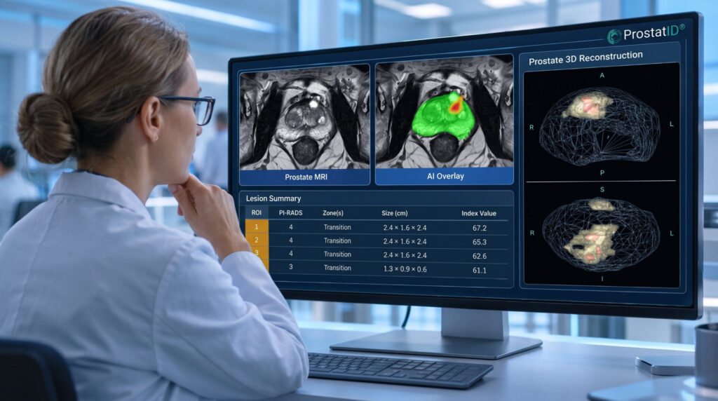
Understanding the critical differences in cancer detection, lesion characterization, and clinical performance—and what makes ProstatID® stand out.
By Randall W. Jones, D.E. (PhD, MBA)
Founder and CEO, Bot Image
Artificial intelligence is rapidly transforming medical imaging, and prostate MRI is no exception. An increasing number of software companies now offer AI tools designed to assist radiologists in interpreting prostate examinations. Many prominently advertise that their products are FDA-cleared.
For radiologists, urologists, and healthcare administrators evaluating these technologies, the expanding marketplace presents an important challenge: How do you distinguish genuinely useful clinical AI from software that offers more limited capabilities?
I frequently hear physicians express frustration after evaluating an AI product that failed to deliver the expected clinical value. Often, the problem begins with a misunderstanding of what the software was actually designed—and cleared—to accomplish.
For investors evaluating the medical AI market, the same distinction matters. Regulatory clearance is an important milestone, but the scope of that clearance, the sophistication of the underlying technology, and the quality of its clinical validation ultimately determine how compelling a product may be.
The essential question is what the AI has been cleared to do, how well it performs, and what additional information it provides to physicians.
Those differences are substantial and understanding them begins with the FDA.
1. FDA-Cleared for What? Understanding the Critical Distinction
One of the most common misconceptions about medical AI is that FDA-cleared products offer comparable clinical capabilities.
In reality, FDA clearance is granted for specific intended uses and indications.
Under the FDA’s 510(k) pathway, manufacturers generally must demonstrate that their devices are substantially equivalent to legally marketed predicate devices, supported by appropriate performance and safety evidence. Medical device registration and listing alone do not establish FDA clearance.
For prostate MRI AI, two FDA product classifications illustrate why these distinctions matter.
QIH: Automated Radiological Image Processing Software
Product code QIH, under 21 CFR §892.2050, encompasses automated radiological image-processing and analysis software.
Depending on its cleared indications, this software may perform functions such as prostate gland segmentation, anatomical measurements, volume calculations, and image visualization.
These capabilities can be valuable. Automated prostate volume measurement, for example, can improve workflow efficiency and support clinical assessments.
However, a product cleared to measure or segment the prostate should not automatically be assumed to have clearance to detect or characterize suspicious cancer lesions.
QDQ: Computer-Assisted Detection and Diagnosis
Product code QDQ, under 21 CFR §892.2090, addresses radiological computer-assisted detection and diagnosis software for lesions suspicious for cancer.
Two important capabilities are involved:
CADe (Computer-Assisted Detection) helps physicians identify and localize suspicious abnormalities that warrant closer examination.
CADx (Computer-Assisted Diagnosis) provides additional information to characterize those findings, such as malignancy-likelihood scores or diagnostic classifications.
These functions influence diagnostic interpretation more directly. The applicable FDA special controls therefore address clinical performance testing, including lesion localization and the effect of AI assistance on reader performance.
ProstatID received FDA 510(k) clearance under product code QDQ on July 8, 2022 (K212783).
Its cleared indications specifically include assisting trained radiologists in detecting, assessing, and characterizing prostate abnormalities, including suspicious cancer lesions.
Other prostate MRI products have subsequently received QDQ clearances. Consequently, the product code establishes a useful starting point for comparison, while the specific indications, outputs, and supporting evidence reveal the more meaningful differences.
2. Beyond Detection: What ProstatID Actually Delivers
Consider what a radiologist must accomplish when interpreting a prostate MRI.
The examination requires reviewing multiple imaging sequences, identifying suspicious regions, distinguishing benign findings from potentially significant cancer, assessing lesion extent, and assigning an appropriate PI-RADS risk assessment.
These tasks require substantial expertise, and interpretation can vary between readers.
ProstatID was developed to provide an additional quantitative perspective throughout this process.
Using three standard MRI sequences—T2-weighted (T2W), diffusion-weighted imaging (DWI), and apparent diffusion coefficient (ADC)—ProstatID analyzes the prostate and generates several clinically relevant outputs.
Its FDA-cleared capabilities include:
- Automated identification of suspicious regions.
- Malignancy-likelihood scores.
- Assessment of lesion locations and estimated boundaries.
- Prostate volume measurement.
- Identification of multiple abnormalities.
- Suggested overall PI-RADS examination score.
ProstatID also provides colorized MRI overlays, three-dimensional representations of suspicious lesions within the prostate, and a PDF report summarizing its findings.
The value lies in bringing detection, characterization, spatial information, and visualization together in a single clinical workflow.
Importantly, ProstatID does not require dynamic contrast-enhanced (DCE) images as an algorithm input. It can therefore analyze compatible biparametric MRI (bpMRI) examinations using T2W, DWI, and ADC sequences, including examinations that also contain contrast-enhanced images.
The interpreting physician remains responsible for reviewing all available imaging information, including DCE when acquired, and incorporating the findings into the clinical assessment.
The combination of detection, quantitative characterization, and spatial visualization creates an opportunity to make prostate MRI findings more informative and actionable.
3. The Technology Difference: Finding Information Beyond Visual Interpretation
Human radiologists interpret MRI through anatomical structures, signal intensity, diffusion restriction, and other visible imaging characteristics.
However, MRI images also contain complex quantitative patterns of signal intensity, texture, and spatial relationships that are exceedingly difficult to distinguish through visual interpretation alone. Research by Lay and colleagues at the National Institutes of Health (NIH) demonstrated how machine-learning algorithms can extract and analyze these features to improve automated prostate cancer detection.
This is where ProstatID’s underlying technology becomes particularly interesting.
Building on this foundational NIH research, ProstatID’s architecture combines deep-learning-based anatomical segmentation with a machine-learning classification approach described in published research and a U.S. patent as a boosted parallel random forest ensemble.
The software first identifies the prostate anatomy and then analyzes quantitative imaging features derived from T2W, DWI, and ADC sequences. Its classification models were trained using MRI data that included biopsy-correlated examples of normal, benign, and cancerous tissue.
An important feature of this approach is its ability to evaluate tissue characteristics at the voxel level.
A voxel is the three-dimensional equivalent of a pixel: a small volume of tissue represented within an MRI examination.
Rather than relying exclusively on a single classification for an entire suspicious region, ProstatID analyzes imaging features across individual voxels and generates a spatial map of estimated cancer likelihood.
Turning quantitative information into a visual risk map
ProstatID converts these calculations into translucent color overlays displayed on the original MRI.
The colors communicate relative suspicion:
- Green represents tissue not suspicious of cancer.
- Yellow indicates increasing levels of suspicion.
- Red highlights regions assigned higher estimated likelihood of clinically significant cancer.
These visualizations allow physicians to see how the software’s assessment varies across the prostate and within suspicious regions.
For instance, ProstatID commonly identifies a region of high suspicion (red) encapsulated within a larger region of moderate suspicion (yellow). This visualization provides additional spatial information about where the greatest concern lies, helping physicians identify areas for precision-targeted biopsy and subsequent clinical management.
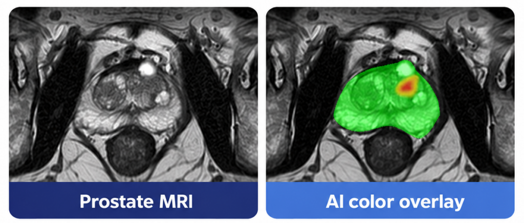
MRI with Color-Coded Overlay from ProstatID
Behind these visualizations is an equally important quantitative assessment. ProstatID assigns each suspicious lesion a continuous risk score, expressed on a 0-100-point scale, that correlates with the probability of clinically significant cancer. Calibrated to the traditional 5-point PI-RADS scale, this score provides finer distinctions in estimated cancer risk than PI-RADS categories alone.
These scores and visualizations represent algorithmic predictions derived from MRI characteristics. As with any AI tool, they do not establish the microscopic presence of cancer cells, which requires pathological confirmation.
Together, the color overlays and quantitative risk scores provide physicians with a more detailed picture of where suspicious tissue is located and how the estimated risk varies within it.
ProstatID transforms complex quantitative imaging patterns into spatially organized information that a physician can readily examine alongside the original MRI.
4. Why Lesion Localization and Boundaries Matter
Identifying a suspicious lesion is an essential first step. Understanding precisely where it is located and how far it may extend presents additional challenges.
This is particularly important when considering MRI-targeted biopsies or evaluating patients who may be candidates for focal treatment.
In routine clinical practice, radiologists estimate lesion boundaries using MRI characteristics. Targeted biopsies can provide pathological confirmation, but when they sample only radiological-selected areas of the prostate, they may be missing cancerous tissues not identified or adequately segmented. Even an accurately targeted biopsy cannot establish the boundaries of an entire tumor.
Detailed correlation between MRI findings and whole-mount pathology following prostatectomy can provide a more comprehensive assessment of tumor extent, but such evidence is not routinely available for patients undergoing initial evaluation.
This is where ProstatID’s spatial risk mapping and three-dimensional lesion visualization provide additional insight. Its FDA-cleared capabilities include assessing the extent of suspicious abnormalities, including lesion boundaries and the identification of multiple lesions.
These outputs can help physicians visualize the estimated distribution of suspicious tissue and understand its relationship to the surrounding prostate anatomy.
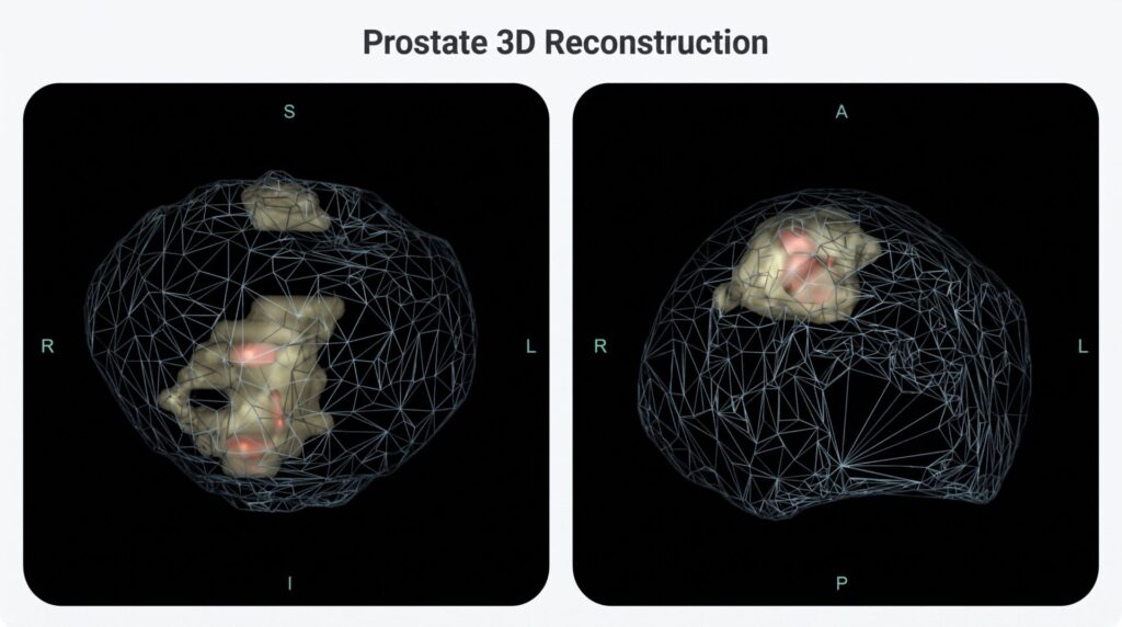
3D Reconstruction from ProstatID
For urologists performing targeted biopsies or evaluating focal treatment options, this information may be valuable as part of a broader clinical assessment.
There is an important distinction, however, between AI-estimated lesion extent and histologically confirmed tumor margins. While detailed spatial risk maps offer valuable imaging-based information, they cannot definitively establish microscopic tumor boundaries.
Further prospective research is needed to establish how these capabilities affect biopsy targeting, treatment decisions, and patient outcomes. Fortunately, researchers are already investigating how AI-generated prostate cancer maps, such as from ProstatID, correlate with targeted biopsy and pathological findings to better understand their potential to improve clinical care.
5. Clinical Evidence: Does ProstatID Improve Physician Performance?
Sophisticated algorithms and impressive visualization are valuable only when they contribute to meaningful clinical performance.
For AI designed to assist radiologists, one particularly important question is:
Do physicians perform better when they use the software?
A useful way to investigate this is through a multi-reader, multi-case (MRMC) study.
In an MRMC study, multiple physicians interpret the same examinations with and without AI assistance. Their performance is evaluated against reference standards, allowing investigators to measure whether AI changes diagnostic accuracy and lesion detection.
ProstatID has undergone this type of clinical assessment.
In a peer-reviewed study published in Current Problems in Diagnostic Radiology in 2024, nine physicians evaluated 150 retrospective biparametric prostate MRI examinations, with and without ProstatID assistance.
The study included MRI examinations from six contributing institutions, representing 12 MRI scanner models, with suspicious findings correlated to biopsy results. The nine physicians independently interpreted the examinations, first without AI assistance and then, following a washout period of at least 30 days, with ProstatID.
Importantly, lesion-detection performance was evaluated against biopsy locations with known pathological findings. This is a particularly rigorous approach because it focuses on detecting and characterizing verified suspicious regions rather than awarding credit for correctly identifying the large volumes of normal tissue throughout the prostate. By evaluating the same physicians using identical examinations and reference standards, the study provides a meaningful comparison of performance with and without AI assistance.
The investigators reported statistically significant improvements in two important measures.
Improved diagnostic discrimination
The reader-averaged area under the receiver operating characteristic curve (ROC AUC) increased from:
0.672 without ProstatID to 0.718 with ProstatID.
That represents an improvement of +0.046 AUC (p = 0.0149).
ROC AUC measures how effectively a diagnostic method distinguishes positive from negative cases across different decision thresholds. Higher values generally indicate better diagnostic discrimination.
Improved lesion-detection performance
The investigators also examined lesion-level detection using free-response ROC analysis, which considers the ability to identify cancerous lesions relative to false-positive findings.
The weighted figure of merit increased from:
0.405 without ProstatID to 0.453 with ProstatID.
That represents an improvement of +0.048 (p = 0.024).
The improvements in ROC AUC and lesion-detection performance are particularly important because they measure the performance of physicians using ProstatID, rather than relying exclusively on the software’s standalone accuracy.
| PUBLISHED CLINICAL EVIDENCE | |
|---|---|
|
Diagnostic discrimination (ROC AUC)
0.672 → 0.718
+0.046 · p = 0.0149
|
Lesion detection (weighted FROC)
0.405 → 0.453
+0.048 · p = 0.024
|
The study also provides another perspective on ProstatID’s detection capabilities through standalone performance analysis. At approximately one false-positive finding per patient, ProstatID achieved 82% sensitivity for detecting clinically significant cancer, compared with approximately 54% for radiologists interpreting the examinations without AI assistance.
Sensitivity vs. Number of False-positives per patient
ProstatID: 0.82, Radiologists alone: 0.54
While this comparison measures the software operating independently rather than its effect on assisted readers, it further illustrates the potential value of ProstatID’s lesion-detection capabilities.
The findings demonstrate statistically significant improvements under the conditions of the study, although performance varied between individual readers. Results from a retrospective study should not be interpreted as guaranteed improvements in every clinical setting.
For healthcare organizations evaluating AI investments, this type of evidence is especially relevant: it directly examines how technology can affect the physicians responsible for interpreting patient examinations.
6. Why Headline AI Accuracy Numbers Can Be Misleading
Medical AI vendors frequently promote sensitivity, specificity, accuracy, or AUC statistics.
These numbers are useful, but comparisons between products require considerable care.
Performance depends on multiple factors, including composition of the patient population, cancer prevalence, lesion characteristics, MRI equipment, image quality, reference standards, and statistical methodology.
Even the definition of successful detection can significantly change the reported result.
Case-level performance evaluates how effectively a system distinguishes patients with and without cancer.
Lesion-level performance evaluates how accurately it identifies specific suspicious regions and correlates those findings with available reference standards.
Similarly, standalone AI performance and AI-assisted physician performance answer different questions.
A standalone AUC measures the algorithm under a particular evaluation method. A reader-study AUC measures physicians interpreting examinations with the technology available to them.
These results should not be directly compared as though they represent the same clinical task.
When evaluating competing technologies, decision-makers should look beyond a single headline statistic and examine:
- The composition of the study population
- The number and experience of participating physicians.
- Whether findings were confirmed by biopsy or surgical pathology.
- The treatment of false positives.
- Whether AI-assisted performance was compared against unassisted interpretation.
They should also consider study funding, authorship, and whether results have undergone peer review.
ProstatID’s published clinical research addresses several of these questions through biopsy-correlated evaluations and controlled reader comparisons.
The most meaningful AI performance claims are those supported by transparent methodology and evidence relevant to the product’s intended clinical use.
7. What Makes ProstatID a Compelling Technology Platform?
The distinctions discussed above have practical implications across the healthcare ecosystem.
For radiologists
ProstatID provides an additional quantitative perspective on suspicious prostate findings.
Its combination of lesion detection, malignancy-likelihood mapping, estimated lesion extent, and suggested PI-RADS assessment can support a more informed interpretation of complex examinations.
The radiologist retains responsibility for the final clinical interpretation.
For urologists
ProstatID’s color overlays and color-coded 3D visualizations provide a way to review suspicious regions, distinguish the most significant risk regions, and their estimated spatial extent.
These outputs may support discussions about targeted biopsies, active surveillance, and treatment planning when considered alongside pathology, PSA, clinical history, and other findings.
For hospitals and imaging centers
The decision to adopt medical AI involves three important considerations: demonstrated clinical performance, integration into existing workflows, and financial viability.
ProstatID addresses workflow efficiency through its cloud-based processing architecture. Once connected to the service, imaging providers can securely transmit an MRI examination for automated analysis. ProstatID performs image quality assessment, detects and characterizes suspicious regions, generates colorized overlays and 3D visualizations, and returns its results for review alongside the original study.
By automating lesion identification, segmentation, and visualization, ProstatID can reduce the manual effort involved in prostate MRI interpretation and preparation for subsequent clinical procedures.
Importantly, ProstatID’s capabilities also align with established insurance reimbursement pathways.
Its advanced 3D reconstruction capabilities may support billing under CPT 76377 when the service meets applicable requirements for medical necessity, physician involvement, interpretation, and documentation. Certain commercial payer policies explicitly recognize 3D prostate segmentation for MRI-ultrasound fusion biopsy preparation.
Another opportunity exists through Category III CPT code 0898T, which addresses prostate cancer estimation mapping derived from fusion-guided biopsy and pathological findings. ProstatID’s 3D mapping capabilities can contribute to this process when combined with the required biopsy and pathology data, margin assessment, and physician interpretation. Coverage and reimbursement for this emerging service vary by payer.
Additionally, the new 2026 Category I CPT codes for prostate biopsy recognize MRI-ultrasound fusion-guided procedures and additional targeted lesions. ProstatID’s lesion localization, 3D segmentation, and RTSTRUCT outputs may facilitate these workflows, providing valuable information to urologists performing targeted biopsies.
Importantly, these reimbursement opportunities have already been demonstrated in clinical practice. Medical practices consulted by Bot Image have reported successfully obtaining insurance reimbursement for qualifying services using applicable CPT codes, including 3D reconstruction, prostate cancer mapping, and fusion-guided biopsy procedures.
These real-world experiences demonstrate how improved workflow efficiency, combined with established reimbursement mechanisms, can support the economic adoption of technologies such as ProstatID. Actual payment amounts and eligibility vary by insurer, clinical application, and care setting.
For healthcare investors
ProstatID brings together several characteristics relevant to evaluating medical AI businesses: FDA-cleared detection and diagnostic-support capabilities, a differentiated technical architecture, published reader-performance evidence, and integration into established clinical workflows.
Its regulatory history is also noteworthy.
ProstatID was cited as a reference device in Siemens Healthineers’ Prostate MR AI FDA submission and identified as a predicate device in DeepHealth ProstateAI’s 2026 submission.
These references demonstrate ProstatID’s established position within the evolving regulatory landscape for prostate MRI AI, although they should not be interpreted as evidence of superiority over those products.
ProstatID’s alignment with existing reimbursable imaging and fusion-biopsy workflows also provides a potential economic incentive for clinical adoption, complementing its demonstrated diagnostic performance and workflow capabilities.
As the market develops, long-term competitive success will depend on continuing clinical validation, physician adoption, workflow compatibility, product development, and demonstrated value to healthcare providers.
8. The Future of Prostate MRI AI Will Be Defined by Clinical Value
The market for prostate MRI AI continues to evolve, and innovation across multiple manufacturers has the potential to improve cancer detection and patient care.
But the presence of an FDA clearance alone tells only part of the story.
The more important questions are what a product has been cleared to do, how thoroughly its performance has been evaluated, and what additional information it provides to clinicians.
ProstatID combines FDA-cleared cancer detection and diagnostic-support capabilities with advanced tissue classification, voxel-level malignancy-likelihood mapping, lesion visualization, and published evidence of improved physician interpretation.
Together, these capabilities reflect Bot Image’s commitment to developing AI that delivers meaningful information for clinical decision-making.
For radiologists, urologists, healthcare administrators, and investors evaluating this rapidly developing field, understanding these differences is essential.
Not all prostate MRI AI is created equal. The technologies that stand apart will be those that demonstrate measurable clinical value—and help physicians make more informed decisions for their patients.
Discover the Difference with ProstatID
Explore how Bot Image’s FDA-cleared prostate MRI AI technology supports cancer detection, lesion characterization, and advanced imaging visualization.
Learn more about ProstatID and request a demonstration
References
FDA clearance and regulatory classifications
U.S. FDA — ProstatID 510(k) Clearance, K212783. FDA decision July 8, 2022. Includes indications for use and 510(k) summary.
U.S. FDA — Product Classification QDQ. Radiological Computer-Assisted Detection/Diagnosis Software.
U.S. FDA — Product Classification QIH. Automated Radiological Image Processing Software.
Clinical research
Maki JH, Patel NU, Ulrich EJ, Dhaouadi J, Jones RW. Part II: Effect of different evaluation methods to the application of a computer-aided prostate MRI detection/diagnosis (CADe/CADx) device on reader performance. Current Problems in Diagnostic Radiology. 2024;53(5):614–623. DOI: 10.1067/j.cpradiol.2024.04.003.
Lay N, Tsehay Y, Greer MD, et al. Detection of prostate cancer in multiparametric MRI using random forest with instance weighting. Journal of Medical Imaging. 2017;4(2):024506.DOI: 10.1117/1.JMI.4.2.024506.
Bot Image — Full-text publication of the MRMC reader study (PDF).
Subsequent FDA submissions
Siemens Healthineers — Prostate MR AI, K241770. Identifies ProstatID as a reference device.
DeepHealth ProstateAI — K253682. Identifies ProstatID as a predicate device.
Patent
Bot Image patent: Dhaouadi J, Ulrich EJ, Jones RW. Automated Prostate Cancer Detection and Diagnosis Using a Boosted Ensemble of Bagging Ensemble Models. U.S. Patent No. 12,626,360, issued May 12, 2026.
Disclosure: Randall W. Jones is a co-author of the cited clinical reader study and is affiliated with Bot Image.
Pioneering Cancer Detection with AI and MRI (and CT)
At Bot Image™ AI, we’re on a mission to revolutionize medical imaging through cutting-edge artificial intelligence technology.
Contact Us