


Blog
How AI Reduces Radiologist Fatigue and Reading Variability in Prostate MRI
The modern radiology reading room is a paradox. It is a place of incredible technological sophistication, where high-definition monitors display the inner workings of the human body in breathtaking detail. Yet, it is also a place of immense human pressure. The silent hum of workstations is often accompanied by the mounting stress of a workload that never seems to end.
Radiologists are the unsung heroes of diagnostic medicine, but they are facing a crisis. The volume of imaging studies has exploded over the last decade, outpacing the number of available specialists. Amidst this deluge, prostate MRI has emerged as a particularly challenging exam. It is a critical tool for detecting one of the most common cancers in men, but it is also one of the most difficult to interpret.
The twin challenges of radiologist fatigue and inter-reader variability are threatening the quality of patient care. When a radiologist is tired, or when two experts disagree on a diagnosis, the patient pays the price in uncertainty, delayed treatment, or unnecessary procedures.
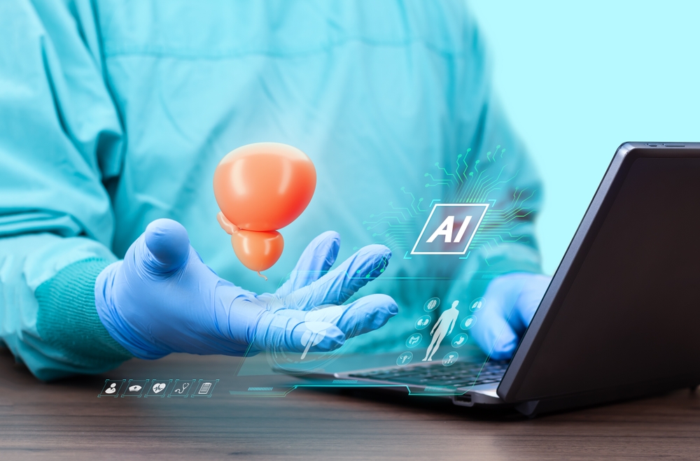
Artificial Intelligence (AI) has arrived not as a replacement for the radiologist, but as a remedy for these very human limitations. Tools like ProstatID™ are revolutionizing the reading room by standardizing interpretation and alleviating the cognitive burden that leads to burnout.
In this comprehensive guide, we will explore the root causes of fatigue and variability in prostate imaging and demonstrate how AI in prostate MRI is creating a more sustainable, accurate future for radiologists and patients alike.
The Cognitive Marathon: Why Radiologists Are Tired
To understand the solution, we must first appreciate the problem. Reading a prostate MRI is not a passive activity; it is an intense cognitive workout.
The Anatomy of an Exam
A standard multiparametric MRI (mpMRI) of the prostate consists of thousands of images. The radiologist must navigate through:
- T2-Weighted Images: To assess anatomy and zonal segmentation.
- Diffusion-Weighted Imaging (DWI): To identify cellular density, a key marker of tumors.
- Apparent Diffusion Coefficient (ADC) Maps: To confirm restricted diffusion.
- Dynamic Contrast-Enhanced (DCE) Sequences: To observe blood flow patterns.
The radiologist isn’t just looking at these images; they are synthesizing them. They must mentally co-register these different sequences in 3D space, compensating for slight patient movements, and filtering out artifacts. They must do this while searching for lesions that can be as small as a few millimeters.
The Volume Problem
Now, multiply that complexity by volume. A radiologist might be expected to read 20, 30, or even 50 complex cases in a single shift. This relentless pace leads to “decision fatigue.” As the day wears on, the brain’s ability to make sharp, high-stakes decisions degrades.
Studies have shown that diagnostic accuracy can dip significantly toward the end of a long shift. This isn’t a failure of skill; it’s a physiological reality of the human brain. We are not built to maintain peak focus for 10 hours straight without aid.
The “Search” vs. “Decide” Imbalance
A significant portion of a radiologist’s time is spent on “search” tasks—scrolling through slices, measuring the gland, and hunting for abnormalities. This is the “grunt work” of radiology. It consumes mental energy that should be reserved for the “decide” tasks—interpreting the findings and formulating a diagnosis. By the time the radiologist finds the lesion, they have already expended valuable cognitive fuel.
The Variability Crisis: When Experts Disagree
If fatigue is the enemy of endurance, variability is the enemy of consistency. One of the most frustrating aspects of prostate MRI is that it is subjective.
The Subjectivity of PI-RADS
The Prostate Imaging Reporting and Data System (PI-RADS) was developed to standardize how prostate MRI is reported. It assigns a score from 1 (highly unlikely to be cancer) to 5 (highly likely to be clinically significant cancer).
While PI-RADS is a massive step forward, it hasn’t solved the problem of subjectivity.
- Reader Experience: An academic radiologist who reads 1,000 prostate MRIs a year will interpret a scan differently than a general radiologist in a community hospital who reads 50.
- The “Eye of the Beholder”: What looks like a PI-RADS 3 (indeterminate) to one doctor might look like a PI-RADS 4 to another.
- Benign Mimics: Conditions like Benign Prostatic Hyperplasia (BPH) or prostatitis can mimic cancer patterns, leading to disagreements on whether a biopsy is necessary.
The Cost of Variability
Reducing variability in radiology is not just an academic exercise; it has real-world consequences.
- Over-diagnosis: If a radiologist is too aggressive (calling many PI-RADS 4s), patients undergo unnecessary biopsies, which carry risks of infection and side effects.
- Under-diagnosis: If a radiologist is too conservative (calling lesions PI-RADS 2), significant cancers may be missed, delaying life-saving treatment.
Patients shouldn’t have to wonder if their diagnosis depends on who read their scan or when it was read. They deserve a consistent standard of care.
Enter AI: The tireless Assistant
This is where Artificial Intelligence steps in. AI does not get tired. It does not have a bad day. It does not get distracted by a phone call. It applies the exact same rigorous mathematical analysis to the first case of the day as it does to the last.
ProstatID™ utilizes deep learning algorithms trained on thousands of biopsy-confirmed cases. It brings a level of consistency and stamina that is impossible for a human to match alone.
How AI Combats Fatigue
ProstatID benefits the tired radiologist by automating the low-value, high-effort tasks.
1. Automated Segmentation
Traditionally, a radiologist must manually outline the prostate gland to calculate its volume (a critical metric for PSA density). This involves clicking, dragging, and adjusting calipers on multiple slices. It is tedious and repetitive.
ProstatID does this instantly. It segments the gland and calculates the volume automatically. By removing this manual labor, the radiologist saves physical clicks and mental energy on every single case.
2. Pre-Identification of Lesions
Instead of starting with a blank screen and searching for a needle in a haystack, the radiologist opens the case to find the “needles” already highlighted. The AI overlays color-coded maps on the T2 images, pointing out suspicious areas.
This changes the workflow from “Search and Detect” to “Verify and Interpret.” The radiologist can immediately focus their attention on the regions of interest identified by the AI. This “head start” drastically reduces the cognitive load required to initiate the read.
3. Decision Support
When a radiologist is fatigued, doubt creeps in. “Is this a real lesion, or just noise?”
ProstatID provides an objective risk score for each detected lesion. This acts as a second opinion. If the AI confirms the radiologist’s suspicion, it provides the confidence to sign the report quickly. If the AI flags something the radiologist missed, it triggers a safety check.
How AI Reduces Variability
By introducing an objective, mathematical standard into the reading process, AI narrows the gap between readers.
1. Standardization of Interpretation
The AI doesn’t “think” like a human; it recognizes patterns based on data. It has “seen” more cases of prostate cancer than any single human could see in ten lifetimes.
When a general radiologist uses ProstatID, they are effectively borrowing the expertise of those thousands of training cases. The AI guides them toward a standard interpretation. Studies have shown that when less experienced readers use AI assistance, their accuracy improves to match that of sub-specialist experts.
2. Consistent Risk Scoring
The AI assigns risk scores based on pixel-level data, not gut feeling. This helps align the PI-RADS scoring across the department. If the AI consistently scores a certain pattern as high-risk, it trains the radiologists to recognize that pattern uniformly. Over time, the entire group moves toward a more cohesive diagnostic standard.
3. Reducing “Satisfaction of Search”
A common source of variability is the “satisfaction of search” error—finding one lesion and stopping, assuming the job is done. The AI scans the entire gland every time. It will flag multiple lesions if they exist. This ensures that a secondary cancer isn’t missed just because the primary one was obvious.
The Human Impact: Beyond the Reading Room
While we focus on the technical benefits of AI in prostate MRI, the ultimate goal is to improve human lives.
For the Radiologist: A Better Work-Life Balance
Burnout is driving radiologists out of the profession early. By reducing the mental grind of the job, AI can help preserve the workforce. A radiologist who finishes their shift feeling less exhausted is a radiologist who is less likely to make errors and more likely to enjoy their career.
For the Patient: Trust and Speed
Patients sense uncertainty. When a doctor says, “We need a second opinion on this scan,” or “The results are inconclusive,” anxiety spikes.
By improving diagnostic confidence and speed, AI helps get definitive answers to patients faster. To understand how these technical improvements translate to patient outcomes, explore our data on how we Discover Our Impact on the clinical journey.
For the Caregiver: Clarity
Behind every patient is a family. The confusion caused by variable diagnoses or missed findings affects them deeply. A clear, accurate diagnosis allows families to plan. For resources on navigating this journey, we have created a dedicated page for caregivers.
The Science of Trust: Why Radiologists are Adopting AI
Adopting new technology requires trust. Radiologists are naturally skeptical of “black boxes.” They need to know that the tool is reliable.
Validated Performance
ProstatID is FDA-cleared. This means it has undergone rigorous testing to prove its safety and efficacy. In clinical validation studies, the use of ProstatID was shown to improve reader performance significantly. It increased sensitivity (finding cancer) and specificity (ruling out non-cancer).
Ground Truth Training
Unlike some AI trained only on radiologist reports (which contain errors), ProstatID was trained on pathology-verified ground truth. The algorithm learned from biopsy results. It learned to see the microscopic truth of the disease. This gives radiologists confidence that the AI is anchoring its findings in biological reality.
Looking Ahead: The Future of Fatigue Management
As imaging technology advances, scans will only get more complex. We are moving toward 3D imaging, radiomics, and personalized medicine. The amount of data per patient will continue to grow.
Without AI, this data growth would be unmanageable for the human brain. With AI, it becomes an opportunity.
We are building a future where the radiologist is the conductor of an orchestra of data, using AI to harmonize the inputs into a clear, precise diagnosis.
This vision extends beyond detection. It moves into treatment planning and therapy guidance. To see where we are taking this technology next, read about our vision for Beyond Detection: How ProstatID™ Aids in Treatment Planning.
Conclusion: A Partnership for Precision
The narrative of “AI vs. Doctor” is false. The future is “AI + Doctor.”
By addressing the critical issues of radiologist fatigue and inter-reader variability, AI is securing the foundation of diagnostic imaging. It is making the reading room a safer, more efficient, and more consistent environment.
When we remove the noise of fatigue and the fog of subjectivity, what is left is clarity. And clarity is what saves lives.
ProstatID benefits are available today. It is not a futuristic concept; it is a practical tool ready to support your practice.
Ready to reduce burnout and improve accuracy in your imaging center? Learn more about the team behind this innovation at About Bot Image, Inc.
Pioneering Cancer Detection with AI and MRI (and CT)
At Bot Image™ AI, we’re on a mission to revolutionize medical imaging through cutting-edge artificial intelligence technology.
Contact Us
